Knee Anatomy
The knee joint is one of the largest joints in the body, and as such, very important in helping us move. If a problem occurs with the knee, it can be very painful and make mobility difficult or impossible. The knee joint is also very complex. Bones, ligaments, tendons, and cartilage work together to provide stability and movement while holding the joint together.
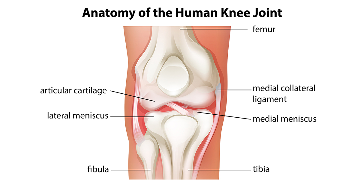
Bones In The Knee
The femur, or thigh bone, is above the knee. Of the bones in the body, the femur is the heaviest, longest, and largest. The tibia, or shin bone, is below the knee. It is the second longest bone in the body. A long thin bone called the fibula is attached to the tibia. The rounded ends of the femur and tibia are called condyle. These boney projections are covered with a material that allows for ease of movement. The patella, or knee cap, is a bone connected by a ligament and a tendon to the knee joint. It is smooth underneath and provides a gliding surface at the outside of the knee joint for ease of movement. It also works to protect the knee joint.
Soft Tissue
The meniscus is a C-shaped piece of cartilage in the knee. There are two menisci in each knee joint, the medial meniscus on the inner side and the lateral meniscus on the outer side the knee joint. These provides stability and protection to the knee while distributing the femur's weight. Articular cartilage is the smooth tissue covering the condyle surfaces where bones meet. This smooth surface serves to provide as little friction between the bones as possible, allowing for ease of movement.
Ligaments are tissue that connect and hold bones together to provide stability in the knee. Tendon are tissue that connect bone to muscle. The anterior cruciate ligament, or ACL, is one of four primary ligaments that hold the knee together. It is located inside the knee joint, running diagonally in the middle of the knee and connecting the bottom of the thigh bone, or femur, to the top of the lower leg bone, or tibia. It is in charge of rotational stability in the knee, but it also prevents the tibia from sliding forward in front of the femur. The posterior cruciate ligament, or PCL, is located behind the ACL. It keeps the tibia from moving backwards. The ACL is injured more often than the PCL, which is stronger. These ligaments work together to create back and forth knee movement.
The medial, or inside, and lateral, or outside, collateral ligaments help to protect the knee from unusual or side-to-side movements. They also serve to stabilize the knee. The patella ligament connects the bottom of the patella to the top of the tibia. The quadriceps tendon connects the top patella to the quadriceps femoral muscle above it. This muscle and tendon combination helps keep the knee cap and knee straight.