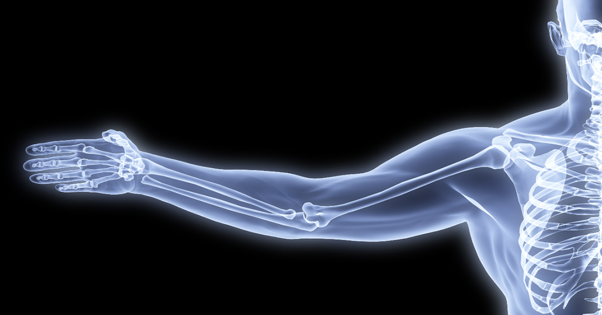
Bone Densitometry, also called dual-energy X-ray absorptiometry (DEXA), is a procedure widely known as a bone scan. It's used to diagnose osteoporosis and to assess the risk of developing fractures. DEXA is simple, quick and noninvasive.
What is a Bone Scan?
Bone scan is the everyday term for bone densitometry, which is formally known as dual-energy X-ray absorptiometry or DEXA. It's a non-invasive diagnostic test that helps to determine the health of your bones. It is useful for detecting the presence of any deformities, and is the standard diagnostic imaging procedure for evaluating osteoporosis. DEXA scans are quick and painless, and they're usually performed on an outpatient basis.
How is a Bone Scan used?
The usual reason your doctor might order a bone scan is to diagnose osteoporosis - a thinning of bone material that can make your bones fragile and more prone to fractures. It's sometimes used to confirm osteoporosis in a person who has other risk factors including age, weight, medical and family history and smoking. It's also a means of tracking the progress of treatment for osteoporosis and other bone loss disorders. It can also be used to confirm the progression of metastatic cancer from bone to surrounding tissue.
What does a DEXA machine look like?
The machine most often used for measuring bone density in hips or spine is called a Central DEXA device. These are usually located in hospitals and medical offices. Central devices have a large, flat table for the patient, with the X-ray imaging unit suspended overhead.
Peripheral, or pDEXA devices are much smaller, and sometimes located in pharmacies and on mobile health vans in the community. A box-shaped structure with an opening for the foot or forearm, the pDEXA device measures bone density in the wrist, heel or finger.
How should I prepare for my bone scan?
When discussing the possibility of a bone scan, be sure to tell your physician your physician if you have recently had a barium examination or have if you have been injected with a contrast material for a computed tomography (CT) scan or radioisotope scan. If you have contrast materials in your system, your doctor might recommend that you wait 10 to 14 days for those to leave your body, before having your DEXA test.
Ahead of the test, you'll be asked a few background questions to ensure that the test is safe to perform.
On the day of the exam you may eat normally. You should not take calcium supplements for at least 24 hours before your exam. Dress comfortably in loose clothing, avoiding zippers, belts or metal buttons. You may be asked to remove some of your clothes and to wear a gown during the exam. You may also be asked to remove jewelry, dental appliances, eyeglasses and other metal objects or clothing that might interfere with the x-ray images. Leave your keys, wallet, phone and pocket change out of the room as well.
Women should always inform their physician and x-ray technologist if there is any possibility that they are pregnant, to avoid exposing the fetus to radiation. If necessary, precautions will be taken to minimize radiation exposure to the baby.
How is the Test Performed?
If the purpose of the scan is to visualize the progress of a cancer, a tracer dye will be injected into your bloodstream before the scan. If any tracer chemical is used, there could be a wait while it moves through your body, so that it will show in the scan.
If the object of the central DEXA examination is to assess bone density in the hip and spine, you'll lie on a padded table that has an x-ray generator is located underneath and an imaging device positioned above you. To assess the spine, your legs are supported on a padded box to flatten the pelvis and lower spine. To assess the hip, the technician will place your foot in a brace that rotates the hip inward. The detector will pass slowly over the area, generating images on a computer monitor.
You must hold very still and may be asked to hold your breath for a few seconds while the detector is moving, to reduce the possibility of a blurred image. The technologist will walk behind a wall or into the next room to activate the x-ray machine.
Peripheral tests are even more simple. The finger, hand, forearm or foot is placed in a small device that obtains a bone density reading within a few minutes.
At many centers a procedure called Lateral Vertebral Assessment (LVA) is now recommended. Performed on the same central DEXA machine, LVA is a low-dose x-ray examination of the spine to screen for vertebral fractures. The LVA test adds a few minutes to the procedure.
Your bone density test will last 10 to 30 minutes, depending on the equipment used and the parts of your body being examined.
Results Obtained
Your results will be rendered as a numerical score:
The T score is used to estimate your risk of developing a fracture. It shows the amount of bone you have compared with a young adult of the same gender with peak bone mass. A score above -1 is considered normal. A score between -1 and -2.5 is classified as osteopenia (low bone mass). A score below -2.5 indicates osteoporosis.
Z score reflects the amount of bone you have compared with other people in your age group and of the same size and gender. If this score is unusually high or low, it may indicate a need for further medical tests.
Your doctor will review with you the results of the scan, and discuss what measure's if any, are indicated by the scores.
Risks of the procedure
Bone scans are very safe. There is a small chance of developing an allergic reaction to any marker dye; this can be treated easily. The radiation exposure is extremely low: less than one-tenth the dose of a standard chest x-ray, and below than a day's exposure to natural radiation.


