What is Autologous Chondrocyte Transplantation?
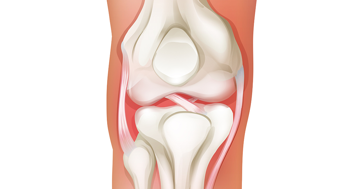
Autologous chondrocyte transplantation is a procedure used to replace damaged cartilage in the knee joint with healthy cartilage cells harvested from the patient. This procedure usually involves two separate surgeries. The first surgery is performed to harvest healthy cartilage cells from the knee joint. Once the cells have been harvested, they are sent off to be cultured in a lab. The second surgery is then performed to implant the lab cultured material into the knee joint.
Who needs Autologous Chondrocyte Transplantation?
This procedure is performed to help treat damaged knee cartilage. Causes of cartilage damage vary. This damaged cartilage may be caused by injury, trauma, or degenerative or other conditions.
What are the steps in Autologous Chondrocyte Transplantation?
Preparing for the Procedure
Anesthesia is administered once the patient has been positioned to allow the surgeon access to the front of the knee, which has been made clean and sterile. Local anesthesia is used at the incision site, and the patient is given general anesthesia or sedation as well.
Accessing the Knee Joint
The surgeon makes two to five small incisions in the knee. Surgical tools are then inserted, including an arthroscopic camera. The arthroscopic camera is used to provide image guidance during the procedure by displaying images from the inside of the knee joint on a monitor in the operating room.
Harvesting the Cartilage Cells
Healthy cartilage cells are then collected from the knee joint. The first surgery is completed and all tools are removed, and the incisions are closed with sutures or surgical staples. The healthy cells are sent to a laboratory, where they are used to grow new cartilage cells. This growth process generally takes four to six weeks. Once the growth process is complete, the patient returns for a second surgery.
Preparing for Implantation
General anesthesia is administered after the patient has been positioned to allow the surgeon access to the front of the knee, which has been made clean and sterile.
Harvesting a Patch
The surgeon makes an incision on the front of the knee to gain access to the damaged cartilage area. Another incision is also made at the area at the top of the lower leg bone. The surgeon harvests a small patch of tissue called periosteum from an area of tissue covering the tibial bone. This harvested tissue is called a periosteal patch.
Applying the Patch
The harvested periosteal patch is placed and attached over the damaged cartilage area. The surgeon then injects underneath the patch the cultured, healthy cells from the first procedure.
Ending the Procedure
The surgeon then closes the incisions with surgical staples or sutures and bandages the knee and leg. The patient must stay at the hospital for observation and physical therapy for one to two days.
After Surgery
It's important that weight is not put on the leg for six to eight weeks. During recovery, patients should adhere to any physical therapy exercises prescribed. While recovery time can vary depending on a variety of a factors including the amount of cartilage damage and the patient's general health, but full recovery time from this procedure can take up to a year.