What is OATS Cartilage Repair Surgery?
OATS cartilage repair surgery, also called mosaicplasty, is a minimally invasive procedure performed to repair damaged cartilage. This is accomplished by removing and replacing damaged cartilage with a graft of healthy cartilage harvested from the patient. This procedure is performed as minimally invasive as possible. Your surgeon will perform the procedure through small incisions using special surgical instruments and tools. A very small camera called an arthroscope is inserted into the joint and used to provide image guidance during the procedure.
Who needs OATS Cartilage Repair Surgery?
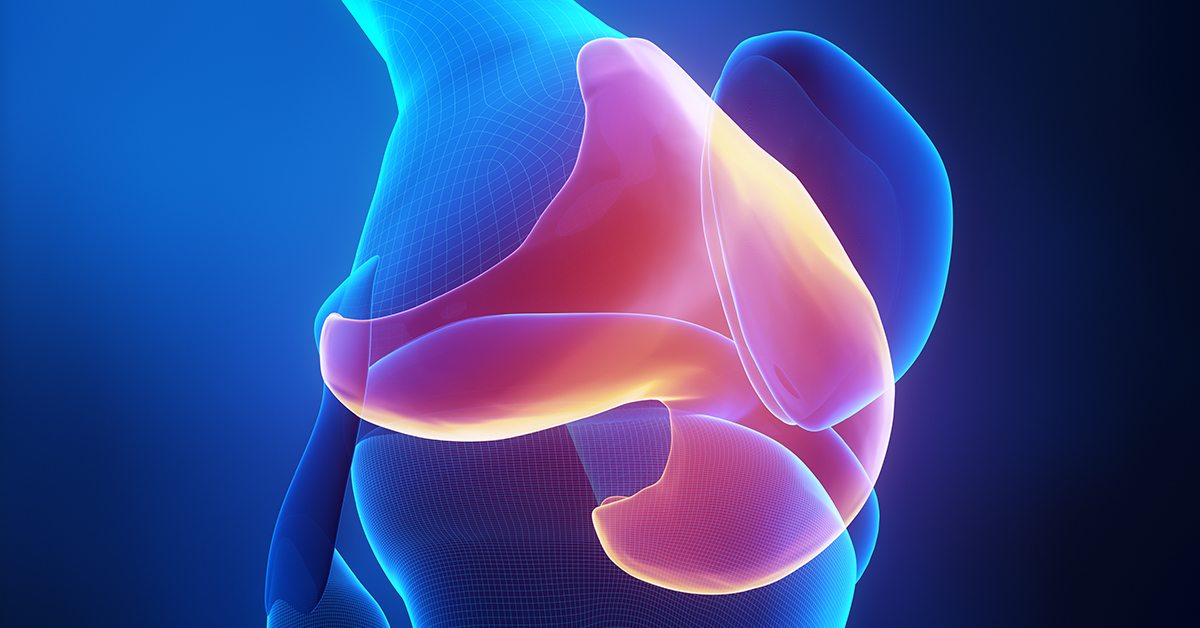
The ends of the bones that meet to make the knee joint are covered in articular cartilage, a smooth tissue that serves to provide a low friction surface for the structures in the knee. This smooth surface can be made rough if damaged, which can be the result of a number of conditions, including trauma, degenerative conditions like arthritis, and others. Symptoms of damaged cartilage in the knee can include joint pain, stability problems like the knee "giving," and popping or locking.
What are the steps in OATS Cartilage Repair Surgery?
Removal of Healthy Cartilage
The surgeon harvests a section of healthy cartilage from a non-weightbearing area in the joint. When selecting the healthy cartilage to be used as a graft, the surgeon considers the size, location, and curvature of the damaged cartilage area to make certain the graft will fit smoothly.
Removal of Diseased Cartilage
The surgeon then removes the area of diseased cartilage, leaving behind a space in which the healthy cartilage can be placed.
Insertion of the Graft
The surgeon then inserts the health graft into the socket created in the previous step, making certain that the graft and surrounding cartilage form a smooth and level surface.
Multiple Grafts
Depending on the size of the damaged area, multiple grafts may need to be used. Each graft is carefully and individually placed. If necessary, allograft may be used to fill larger damaged areas.
End of Procedure
The bone from the damaged area may be used to fill the hole created at the donor site, but the surgeon may decide to leave the donor area open to heal.
After Surgery
Rehabilitation is important in ensuring the procedure's success after the surgery. Crutches may be needed during for up to two months after surgery. Physical therapy and exercise will be prescribed in order to help return normal weight bearing and motion range.